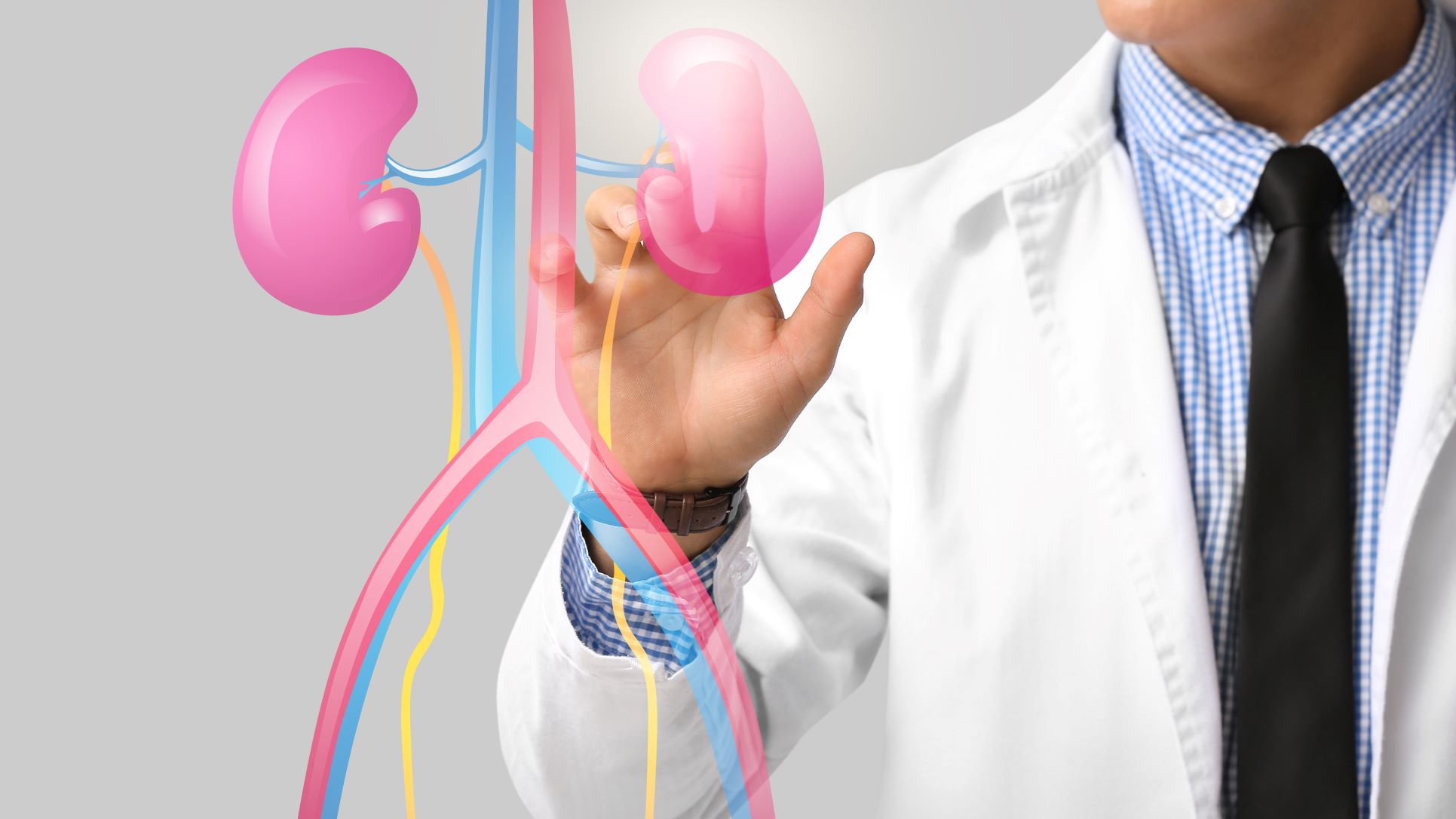
The term left renal concretion is often used in an imaging report to describe a small mineralized deposit or calculus (kidney stone) located in the left kidney.
A renal calculus forms when minerals and salts in urine become concentrated and crystallize. Kidney stones can vary considerably in size, composition and location. Treatment is therefore not determined simply by the word “concretion” on a scan. The report needs to be interpreted together with the stone's size, exact location, symptoms, obstruction, infection status and kidney function.
A small left renal concretion that is not causing symptoms may require monitoring rather than immediate removal. A larger or obstructing stone may need active treatment.
At Prabhakar Bhurke Clinic, Dr. Sandip Prabhakar Bhurke, super specialist nephrologist and transplant physician, provides kidney evaluation and can assess kidney-function concerns associated with renal stones and coordinate appropriate specialist treatment when stone removal is required.
Does Left Renal Concretion Always Mean a Kidney Stone?
In many imaging reports, “renal concretion” is used to describe a calcified focus consistent with a kidney stone.
However, the exact meaning depends on the imaging modality and radiologist's report.
A doctor may need to determine:
-
Whether the finding is definitely a stone
-
Its measurement
-
Whether it is located inside a calyx or renal pelvis
-
Whether there are multiple stones
-
Whether the stone is obstructing urine flow
-
Whether there is associated hydronephrosis
-
Whether infection is present
This is important because not every small calcification seen near the kidney necessarily requires a stone-removal procedure.
What Causes a Left Kidney Concretion?
Kidney stones can develop when certain substances in urine become sufficiently concentrated to form crystals.
Factors associated with stone formation include:
-
Low fluid intake
-
High urinary calcium
-
High urinary oxalate
-
High uric acid
-
Certain metabolic disorders
-
Recurrent urinary infections
-
Family history
-
Obesity
-
Some medications
-
Certain dietary patterns
-
Conditions such as hyperparathyroidism or renal tubular acidosis
The exact cause depends on the individual and the type of stone.
What Symptoms Can a Left Renal Concretion Cause?
A small stone remaining inside the kidney may cause no symptoms at all.
Symptoms are more likely when a stone moves or interferes with urine drainage.
Possible symptoms include:
-
Pain on the left side or flank
-
Pain below the ribs
-
Pain radiating toward the lower abdomen or groin
-
Blood in the urine
-
Burning while urinating
-
Frequent urge to urinate
-
Nausea
-
Vomiting
-
Cloudy or unpleasant-smelling urine
A stone that blocks urine flow can produce significant pain and may affect kidney function if obstruction persists.
Can a Left Renal Concretion Be Harmless?
Yes, depending on its size, position and clinical circumstances.
Some small, non-obstructing kidney stones are discovered incidentally during ultrasound or CT performed for another reason.
If the stone is stable and the patient has no significant symptoms, the doctor may recommend observation with appropriate follow-up imaging.
However, surveillance is not the same as ignoring the finding. Growth, migration, recurrent infection, obstruction or symptoms can change the treatment plan.
How Is a Left Renal Concretion Diagnosed?
The treatment decision starts with understanding exactly what the imaging shows.
Ultrasound
Ultrasound can identify many kidney stones and may also demonstrate swelling of the kidney or urinary obstruction.
It is commonly used because it does not involve radiation.
CT Scan
A non-contrast CT scan can provide detailed information about:
-
Stone size
-
Stone location
-
Number of stones
-
Stone density
-
Urinary obstruction
-
Other urinary tract findings
CT may be particularly useful when the diagnosis or treatment plan remains uncertain.
Urine Examination
Urinalysis can help identify:
-
Blood
-
Infection
-
Crystals
-
Other abnormalities
A urine culture may be recommended when infection is suspected.
Blood Tests
Depending on the situation, testing may include:
-
Serum creatinine
-
eGFR
-
Electrolytes
-
Calcium
-
Uric acid
-
Blood count
-
Other tests based on suspected causes
Kidney-function assessment becomes particularly important when obstruction, infection or multiple stones are present.
When Does a Left Renal Concretion Need Treatment?
Not every renal concretion requires immediate removal.
A doctor may consider active treatment when the stone:
-
Causes recurrent or significant pain
-
Obstructs urine flow
-
Is associated with infection
-
Increases in size
-
Causes recurrent urinary problems
-
Is unlikely to pass or resolve spontaneously
-
Threatens kidney function
-
Is sufficiently large or located in a way that makes observation unsuitable
Treatment decisions are individualized according to stone characteristics and the patient's overall health.
Can a Small Left Renal Concretion Pass Naturally?
Some small kidney stones can pass naturally, particularly when they leave the kidney and enter the ureter.
However, the likelihood of passage depends strongly on stone size and location.
A stone that remains inside the kidney is not necessarily on a path to pass immediately.
Patients should therefore not assume that every stone seen on a scan will simply pass with water or medication.
Left Renal Concretion Treatment Options
The appropriate treatment depends on the individual stone.
Observation
For selected small, asymptomatic and non-obstructing stones, active surveillance may be reasonable.
Follow-up imaging can determine whether the stone remains stable or changes over time.
Medical Management
Medicines may be used for:
-
Pain control
-
Selected stones that are likely to pass
-
Treatment of certain metabolic abnormalities
-
Prevention of recurrent stones
Most established kidney stones cannot simply be dissolved with a general “stone medicine.” Certain uric acid stones may be amenable to medically supervised dissolution therapy.
Shock Wave Lithotripsy
Shock wave lithotripsy (SWL/ESWL) uses externally generated shock waves to fragment a stone into smaller pieces.
The suitability of SWL depends on factors such as:
-
Stone size
-
Location
-
Composition or density
-
Kidney anatomy
-
Patient characteristics
Fragments may then pass through the urinary tract.
Ureteroscopy and Laser Lithotripsy
With ureteroscopy, a thin instrument is passed through the urinary tract to reach the stone.
Laser energy can fragment the stone, and selected fragments may be removed using a small basket.
A temporary ureteral stent may be placed in some cases to support urine drainage after the procedure.
RIRS
Retrograde intrarenal surgery (RIRS) is a flexible ureteroscopic approach used to reach stones located inside the kidney.
A laser can fragment the stone into smaller pieces.
RIRS may be considered for selected renal stones depending on their size, location and anatomy.
PCNL
Percutaneous nephrolithotomy (PCNL) involves creating a small access tract through the skin directly into the kidney.
Specialized instruments are then used to break and remove the stone.
PCNL is particularly useful for larger renal stone burdens and selected complex stones.
Which Treatment Is Best for a Left Renal Concretion?
There is no single procedure that is best for every left renal concretion.
The decision may depend on:
| Factor | Why it matters |
|---|---|
| Stone size | Larger stones may be less suitable for simple observation |
| Location | Different kidney locations have different treatment considerations |
| Density | Harder stones may respond differently to SWL |
| Symptoms | Persistent pain may favor active treatment |
| Obstruction | Blockage may require prompt intervention |
| Infection | Infection may change the timing and type of treatment |
| Kidney function | Reduced function may require additional precautions |
| Anatomy | Kidney structure can influence procedural choice |
| Previous treatment | Prior procedures may affect the next approach |
A specialist should review the actual imaging rather than selecting treatment from stone size alone.
Can Drinking More Water Remove a Left Renal Concretion?
Adequate fluid intake is important for preventing future kidney stones, but drinking excessive water does not reliably remove an established renal concretion.
If a stone is obstructing urine flow, forcing large quantities of water is not a substitute for medical treatment.
For people with kidney failure, heart disease or prescribed fluid restrictions, fluid intake must be individualized.
For stone prevention, adequate fluid intake helps dilute urine, but the appropriate amount should be discussed with a healthcare professional.
What If the Left Renal Concretion Causes an Infection?
A kidney stone associated with urinary infection requires careful evaluation.
Warning signs include:
-
Fever
-
Chills
-
Flank pain
-
Burning urination
-
Cloudy or foul-smelling urine
-
Vomiting
-
Feeling severely unwell
An infected obstructed urinary tract can become a medical emergency.
In such circumstances, the immediate priority may be restoring urine drainage, followed by definitive stone treatment once the infection has been appropriately managed.
Do not delay medical attention if fever or chills occur with known urinary obstruction.
Can a Left Renal Concretion Damage Kidney Function?
A small, non-obstructing stone does not necessarily cause kidney damage.
However, persistent obstruction, recurrent infection or extensive stone disease can potentially impair kidney function.
Kidney function may be assessed using:
-
Creatinine
-
eGFR
-
Urine testing
-
Blood pressure
-
Imaging when indicated
Patients with a solitary functioning kidney, pre-existing CKD or significant obstruction may require particularly careful evaluation.
What Happens After Left Kidney Stone Removal?
Follow-up depends on the treatment performed and the patient's risk of recurrence.
The doctor may recommend:
-
Follow-up imaging
-
Urine testing
-
Stone analysis
-
Blood tests
-
Metabolic evaluation
-
Dietary changes
-
Preventive medication when indicated
If the stone is retrieved, laboratory analysis can help identify its composition. In selected patients, a 24-hour urine collection can help identify factors contributing to recurrent stone formation.
How Can Future Left Kidney Stones Be Prevented?
Prevention should be based on the type of stone and the patient's risk factors.
General measures may include:
Maintain Appropriate Fluid Intake
Adequate fluid helps dilute urine and may reduce the concentration of stone-forming substances.
Reduce Excess Sodium
High sodium intake can increase the risk of some calcium-containing kidney stones.
Follow an Individualized Diet
Depending on stone type, a doctor or dietitian may recommend adjustments to sodium, animal protein, oxalate or other dietary factors.
Do Not Unnecessarily Avoid Calcium
For many calcium stone formers, dietary calcium should not simply be eliminated without professional advice. The correct dietary approach depends on the type of stone and overall health.
Treat Recurrent Infections
If infection-related stones are suspected, appropriate diagnosis and treatment of urinary infection are important.
Complete Metabolic Evaluation When Appropriate
People with recurrent or high-risk stones may benefit from additional testing to identify preventable metabolic abnormalities.
When Should You Seek Urgent Medical Care?
A known left renal concretion requires urgent assessment if you develop:
-
Fever or chills
-
Severe or rapidly worsening flank pain
-
Persistent vomiting
-
Very little or no urine
-
Severe weakness
-
Confusion
-
Blood in urine accompanied by worsening symptoms
-
Symptoms of urinary obstruction
Kidney stones can cause urinary blockage, infection and, in some cases, loss of kidney function if complications are not appropriately treated.
Left Renal Concretion Evaluation at Prabhakar Bhurke Clinic
At Prabhakar Bhurke Clinic, Dr. Sandip Prabhakar Bhurke, super specialist nephrologist and transplant physician, provides individualized evaluation for patients with kidney stones and related kidney-function concerns.
Depending on the patient's situation, evaluation may include:
-
Review of ultrasound or CT reports
-
Assessment of stone size and location
-
Kidney-function testing
-
Urine examination
-
Evaluation for urinary infection
-
Assessment of obstruction
-
Review of recurrent stone history
-
Evaluation of metabolic risk factors
-
Kidney-protection strategies
-
Coordination with an appropriate urological specialist when stone removal is required
The treatment approach depends on the actual stone characteristics rather than the term “renal concretion” alone.
Frequently Asked Questions
What does left renal concretion mean?
Left renal concretion commonly refers to a mineralized deposit or kidney stone located in the left kidney. The imaging report should be reviewed to determine its exact size, location and clinical significance.
Is left renal concretion the same as a kidney stone?
Often, the term is used to describe a renal calculus or stone seen on imaging. Your doctor should interpret the finding in the context of the complete scan.
Does a left renal concretion need surgery?
Not always. Small, asymptomatic and non-obstructing stones may sometimes be monitored. Symptomatic, enlarging, obstructing or complicated stones may require active removal.
Can medicine dissolve a left renal concretion?
Most kidney stones cannot be dissolved with ordinary medicines. Certain uric acid stones may respond to medically supervised urine alkalinization.
Can a left renal concretion be removed with laser treatment?
Yes. Selected kidney stones can be treated with flexible ureteroscopy or RIRS using laser lithotripsy. The appropriate procedure depends on stone size, location and other factors.
Is PCNL used for left kidney stones?
Yes. PCNL is an established procedure for removing larger or complex kidney stones. Whether it is appropriate depends on the individual stone and the patient's health.
Can a renal concretion cause kidney pain?
It can, particularly if the stone moves, obstructs urine flow or causes inflammation. However, many small kidney stones remain painless.
Can a left renal concretion affect creatinine?
A small non-obstructing stone may not affect creatinine. Significant obstruction, infection or extensive stone disease can impair kidney function in some patients.
Conclusion
Left renal concretion treatment depends on what the imaging finding represents and, most importantly, the stone's size, location, symptoms, obstruction, infection status and effect on kidney function.
Some small, non-obstructing renal stones can be monitored, while symptomatic or complicated stones may require treatment with SWL, ureteroscopy with laser, RIRS or PCNL.
Drinking an appropriate amount of fluid and making individualized dietary changes can help reduce the risk of future stones, but excessive water intake is not a substitute for treatment of an obstructing stone.
If a left renal concretion is associated with severe pain, fever, chills, reduced urine output or abnormal kidney-function tests, prompt medical assessment is important.
For individualized kidney evaluation and management of renal stone-related kidney concerns, consult Dr. Sandip Prabhakar Bhurke at Prabhakar Bhurke Clinic.
Medical Disclaimer
This article is intended for general educational and informational purposes only and is not a substitute for professional medical diagnosis, treatment or individualized medical advice. “Renal concretion” is an imaging term that may refer to a kidney stone or calcified focus, and the significance of the finding depends on the actual imaging appearance and clinical context. Treatment varies according to stone size, location, composition, symptoms, obstruction, infection, kidney function and overall health. Do not attempt to force a stone to pass by excessive water intake or use unprescribed stone medicines, painkillers or herbal remedies. Fever, chills, severe flank pain, persistent vomiting, very little or no urine, confusion or rapidly worsening symptoms require prompt medical attention. Consult Dr. Sandip Prabhakar Bhurke or another qualified kidney or urinary-stone specialist for an individualized treatment plan.