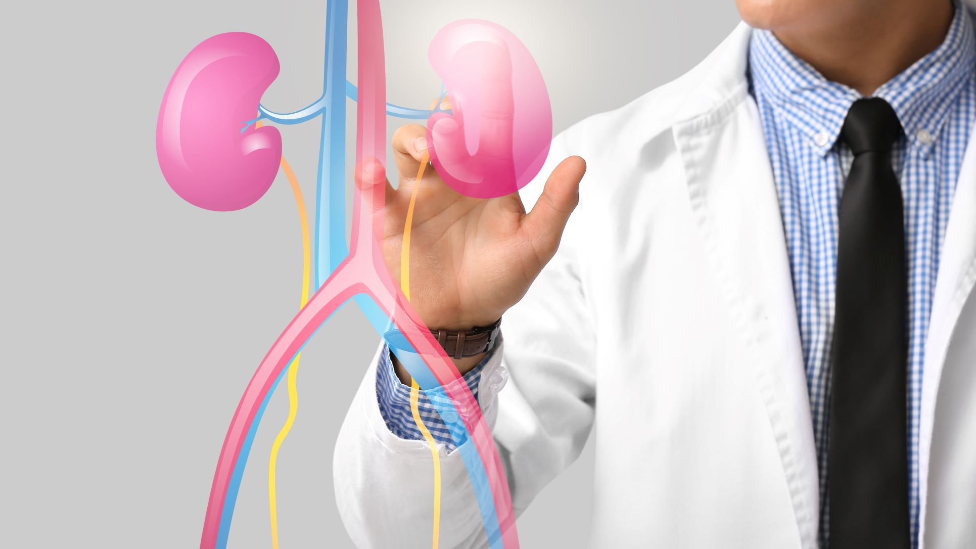
The calyces are parts of the kidney's collecting system. Urine produced by the kidney passes through small collecting spaces called minor calyces before moving into larger calyces and then the renal pelvis.
A kidney stone in a calyx is therefore a stone located within one of these collecting spaces.
A calyceal stone may remain inside the kidney without causing symptoms for a long period. In other cases, it can grow, move into the renal pelvis or ureter, cause obstruction, contribute to recurrent infection or produce pain and blood in the urine.
The appropriate kidney stone in calyx treatment depends on more than stone size alone. Location, stone density, number of stones, symptoms, obstruction, kidney function, infection risk and the patient's overall health all influence treatment selection.
At Prabhakar Bhurke Clinic, Dr. Sandip Prabhakar Bhurke, super specialist nephrologist and transplant physician, provides specialist evaluation of kidney conditions and kidney-function problems associated with urinary stones.
Why Does a Stone Form in the Kidney Calyx?
Kidney stones develop when certain substances in urine become concentrated enough to form crystals.
Depending on the stone type, these substances may include:
-
Calcium
-
Oxalate
-
Uric acid
-
Phosphate
-
Other stone-forming compounds
Factors that may increase stone risk include:
-
Low fluid intake
-
High dietary sodium
-
Certain dietary patterns
-
Family history of kidney stones
-
Metabolic abnormalities
-
Recurrent urinary infections
-
Some medications
-
Certain inherited disorders
Finding the reason for recurrent stone formation can be important for prevention.
Does the Location of a Calyceal Stone Matter?
Yes.
A stone can be located in the:
-
Upper calyx
-
Middle or interpolar calyx
-
Lower calyx
-
Renal pelvis
The location can influence the likelihood that stone fragments will leave the kidney after treatment.
For example, lower-pole stones can sometimes be more challenging for fragments to clear naturally because of the anatomy and drainage angle of the lower kidney.
Therefore, a treatment that works well for one calyceal stone may not be the most suitable option for another.
What Symptoms Can a Calyceal Kidney Stone Cause?
A stone that remains within the kidney may produce no symptoms.
When symptoms occur, they may include:
-
Dull or sharp pain in the side or back
-
Flank discomfort
-
Blood in urine
-
Nausea
-
Vomiting
-
Urinary discomfort
-
Recurrent urinary infections
A stone that moves from the calyx toward the ureter may produce more severe, colicky pain.
Importantly, the absence of pain does not necessarily mean that a calyceal stone should be ignored. Stone size, growth, obstruction, infection and kidney function should also be considered.
How Is a Calyceal Stone Diagnosed?
The first step is usually to establish the stone's location and characteristics.
Ultrasound
Ultrasound can identify many kidney stones and may show associated hydronephrosis or urinary obstruction.
It is also useful for follow-up in selected patients.
Non-Contrast CT
A non-contrast CT scan can provide detailed information about:
-
Stone size
-
Exact location
-
Number of stones
-
Stone density
-
Urinary obstruction
-
Relevant kidney anatomy
CT findings can help the treating specialist select an appropriate intervention when active treatment is required.
Urine Testing
Urinalysis may identify:
-
Blood
-
Infection
-
Crystals
-
Other abnormalities
A urine culture may be required when infection is suspected.
Blood Tests
Depending on the situation, testing may include:
-
Serum creatinine
-
eGFR
-
Electrolytes
-
Blood count
-
Other metabolic investigations
When Does a Calyceal Kidney Stone Need Treatment?
Not every stone inside a kidney requires immediate removal.
Observation may be considered when a stone is:
-
Small
-
Asymptomatic
-
Not obstructing urine flow
-
Not causing recurrent infection
-
Stable on imaging
-
Not associated with significant kidney-function problems
Active treatment may be considered when there is:
-
Persistent or recurrent pain
-
Stone growth
-
Urinary obstruction
-
Recurrent infection
-
Blood in urine associated with the stone
-
Reduced kidney function attributable to obstruction or complications
-
Significant stone burden
-
A low likelihood of spontaneous passage or clearance
-
Patient-specific reasons for planned removal
The decision should be individualized rather than based only on the fact that a stone is present.
Observation for a Calyceal Stone
A small, incidentally detected stone may sometimes be monitored instead of immediately removed.
Follow-up may involve:
-
Periodic imaging
-
Assessment of symptoms
-
Kidney-function monitoring when appropriate
-
Preventive dietary advice
-
Evaluation for recurrent stone risk
If the stone increases in size or begins causing symptoms, the treatment plan may need to change.
Patients should follow the follow-up schedule recommended by their treating specialist.
Medicines for Kidney Stone in Calyx
Medicines usually do not remove or dissolve most calyceal stones.
Medication may instead be used for:
-
Pain control
-
Treatment of infection when bacterial infection is present
-
Prevention of recurrent stones in selected patients
-
Medical dissolution of certain uric acid stones under specialist supervision
The exact medicine depends on the stone type and the patient's kidney function.
Do not take over-the-counter or herbal stone remedies assuming they will dissolve a calyceal stone.
Can Drinking Water Remove a Calyceal Stone?
Adequate fluid intake is an important part of kidney stone prevention.
However, drinking excessive quantities of water does not reliably remove a stone that is sitting inside a kidney calyx.
If a stone is large or obstructing urine drainage, forcing excessive fluid intake may cause discomfort and does not physically eliminate the obstruction.
Fluid recommendations should be individualized, particularly in people with reduced kidney function, heart disease or other fluid-related conditions.
RIRS for Calyceal Kidney Stones
Retrograde intrarenal surgery (RIRS) is a minimally invasive endoscopic technique that can be used to treat selected kidney stones.
A flexible ureteroscope is passed through the natural urinary tract to reach the kidney and inspect the calyces.
The stone can then be fragmented using laser energy.
Depending on its size and location, fragments may be removed using a basket or broken into small pieces that can pass through the urinary tract.
RIRS can be particularly useful when access to a calyx is feasible and an endoscopic approach is appropriate.
Some larger or complex stone burdens may require staged treatment.
Laser Treatment for a Calyceal Stone
Laser lithotripsy can fragment a kidney stone into smaller pieces.
During flexible ureteroscopy or RIRS:
-
The ureteroscope is advanced into the kidney.
-
The affected calyx is identified.
-
Laser energy is applied to the stone.
-
The stone is fragmented.
-
Selected fragments may be removed.
-
A temporary ureteral stent may be placed when appropriate.
Laser treatment is not automatically the right choice for every calyceal stone. Stone size, location, anatomy and the desired degree of stone clearance all matter.
ESWL for a Calyceal Stone
Extracorporeal shock wave lithotripsy (ESWL or SWL) uses externally generated shock waves to fragment selected kidney stones.
Whether ESWL is appropriate depends on:
-
Stone size
-
Stone location
-
Stone density
-
Kidney anatomy
-
Patient body habitus
-
Urinary drainage
-
Infection status
The resulting fragments must then leave the kidney through the urinary tract.
Lower-pole stones can sometimes have less favorable fragment clearance, so the stone's exact calyceal location should be considered before choosing SWL.
Multiple sessions may occasionally be required.
When Is PCNL Considered?
Percutaneous nephrolithotomy (PCNL) is generally used for larger or more complex renal stone burdens.
Through a small access tract made into the kidney, instruments are introduced to fragment and remove the stone.
PCNL may be considered when:
-
The stone burden is large
-
Stones are complex or multiple
-
Complete clearance is difficult with other techniques
-
The stone anatomy makes another approach less suitable
PCNL is a procedure performed by an appropriate surgical stone specialist, generally within urological care.
For smaller uncomplicated calyceal stones, less invasive approaches may be sufficient depending on the circumstances.
Which Treatment Is Best for a Calyceal Stone?
There is no single treatment that is best for every calyceal stone.
The specialist may compare:
| Treatment | When It May Be Considered |
|---|---|
| Observation | Small, stable and asymptomatic stones in selected patients |
| SWL/ESWL | Selected stones with favorable size, density and location |
| RIRS with laser | Selected renal stones accessible through flexible ureteroscopy |
| PCNL | Larger or complex renal stone burdens |
| Medical treatment | Prevention or selected dissolvable stone types |
The final choice depends on imaging, stone characteristics, symptoms, kidney function and patient-specific factors.
Lower Calyx Stone Treatment
A stone in the lower calyx, also called a lower-pole stone, deserves particular attention because the anatomy may make spontaneous fragment clearance more difficult.
Depending on stone size and other characteristics, treatment may include:
-
Observation
-
SWL in selected cases
-
RIRS with laser
-
PCNL for larger stone burdens
The choice should be based on imaging and the expected likelihood of achieving satisfactory stone clearance.
Can a Calyceal Stone Cause Hydronephrosis?
A stone located entirely within a calyx may not cause significant obstruction.
However, if it moves into the renal pelvis or ureter and blocks urine drainage, hydronephrosis can develop.
Persistent obstruction may affect kidney function.
If imaging shows hydronephrosis together with a stone, the cause and severity of obstruction should be assessed.
Calyceal Stone and Kidney Infection
Kidney stones and urinary infections can sometimes occur together.
A stone may contribute to impaired urinary drainage or recurrent infection.
More concerning is an infected obstructed urinary system, which can become a medical emergency.
Symptoms such as:
-
Fever
-
Chills
-
Severe flank pain
-
Vomiting
-
Reduced urine output
-
Feeling severely unwell
require urgent medical assessment.
When obstruction and infection occur together, urgent drainage may be needed before definitive stone removal.
Does a Calyceal Stone Damage the Kidney?
A small, non-obstructing stone may not cause significant kidney damage.
However, complications can occur when a stone causes:
-
Persistent obstruction
-
Recurrent infection
-
Significant inflammation
-
Repeated urinary tract problems
Patients with known stones and abnormal creatinine or reduced eGFR may require closer kidney-function evaluation.
What Happens Before Calyceal Stone Removal?
Before an intervention, the medical team may assess:
-
Stone size
-
Exact calyceal location
-
Stone density
-
Number of stones
-
Kidney anatomy
-
Kidney function
-
Urine culture
-
Blood tests
-
Current medicines
-
Other health conditions
If a urinary infection is present, it generally needs appropriate treatment before definitive stone removal.
The specialist will also discuss whether a single procedure is likely to be sufficient or whether staged treatment may be necessary.
Recovery After Calyceal Stone Treatment
Recovery depends on the procedure used.
After RIRS, SWL or PCNL, some patients may experience:
-
Mild blood in urine
-
Temporary urinary discomfort
-
Flank soreness
-
Increased urinary frequency
-
Passage of stone fragments
A ureteral stent may be placed after certain procedures.
Follow-up imaging may be recommended to determine whether residual stone fragments remain.
Patients should follow their treating specialist's instructions regarding medicines, activity, fluid intake and stent removal.
How Can Another Calyceal Stone Be Prevented?
Removing a stone does not necessarily remove the reason why it formed.
Prevention may involve:
Adequate Fluid Intake
Maintain sufficient fluid intake according to your doctor's recommendations.
Reduce Excess Sodium
High sodium intake can increase urinary calcium excretion in many people and may contribute to calcium-stone formation.
Identify the Stone Type
Stone analysis can provide useful information about the material that formed the stone.
Metabolic Evaluation
People with recurrent or high-risk stones may benefit from blood tests and, when appropriate, 24-hour urine testing.
Follow Preventive Treatment
Depending on the stone type, a specialist may recommend dietary measures or preventive medicines.
When Should You See a Kidney or Stone Specialist?
Medical evaluation is advisable if imaging shows a calyceal stone, particularly when there is:
-
Recurrent flank pain
-
Stone growth
-
Blood in urine
-
Recurrent urinary infection
-
Hydronephrosis
-
Reduced kidney function
-
Multiple or large stones
-
A history of recurrent stone formation
Because stone removal is generally a urological procedure, patients may require coordinated care between nephrology and urology when kidney function or medical kidney problems are also involved.
Kidney Stone Evaluation at Prabhakar Bhurke Clinic
At Prabhakar Bhurke Clinic, Dr. Sandip Prabhakar Bhurke, super specialist nephrologist and transplant physician, provides individualized evaluation for kidney-function abnormalities and kidney conditions associated with urinary stones.
Depending on the clinical situation, assessment may include:
-
Review of ultrasound or CT reports
-
Kidney-function testing
-
Creatinine and eGFR assessment
-
Evaluation of urinary obstruction
-
Assessment of recurrent urinary infection
-
Review of stone-related kidney complications
-
Preventive kidney-stone counseling
-
Coordination with appropriate urological specialists when a stone-removal procedure is required
The treatment pathway depends on the stone's location, size, density, symptoms, obstruction status and the patient's overall health.
Frequently Asked Questions
What is a kidney stone in the calyx?
It is a stone located within one of the collecting spaces of the kidney called the calyces.
Can a calyceal stone pass naturally?
Some small kidney stones may eventually move into the urinary tract and pass, but a stone remaining inside a calyx may also stay there for a prolonged period. Passage depends on size, location and urinary anatomy.
Does every calyceal stone require surgery?
No. Small, stable and asymptomatic stones may sometimes be monitored. Active treatment is more likely to be considered when stones grow, cause symptoms, infection, obstruction or other complications.
What is the treatment for a lower calyx stone?
Depending on its size and characteristics, treatment may include observation, ESWL, RIRS with laser or PCNL for larger stone burdens.
Can RIRS remove a calyceal stone?
Yes. RIRS can access many parts of the kidney through a flexible ureteroscope and may be used with laser lithotripsy for selected calyceal stones.
Can medicine dissolve a calyceal kidney stone?
Most kidney stones cannot be dissolved with medicine. Selected uric acid stones may be treated with medically supervised urine alkalinization.
Does drinking more water remove a kidney calyx stone?
No. Adequate hydration supports prevention and general urinary health, but excessive water intake does not reliably remove a stone lodged in a kidney calyx.
Can a calyceal stone cause kidney damage?
A stone that causes persistent obstruction or recurrent infection can potentially affect kidney function. Non-obstructing stones may not cause significant damage, but they should be monitored according to the treating specialist's advice.
Conclusion
Kidney stone in calyx treatment depends on where the stone is located, how large and dense it is, whether it is growing, and whether it causes symptoms, obstruction, infection or kidney-function problems.
Small and stable calyceal stones may sometimes be monitored, while active treatment can involve RIRS with laser, ESWL or PCNL, depending on the individual stone characteristics.
Lower-pole stones may require additional consideration because fragment clearance can be affected by kidney anatomy. A stone associated with infection and urinary obstruction requires urgent medical attention.
If a scan has identified a stone in your kidney calyx, do not rely solely on home remedies or excessive water intake. A specialist assessment can help determine whether observation, preventive care or active stone removal is appropriate.
For individualized kidney evaluation and management, consult Dr. Sandip Prabhakar Bhurke at Prabhakar Bhurke Clinic.
Medical Disclaimer
This article is intended for general educational and informational purposes only and should not be considered a substitute for professional medical advice, diagnosis or treatment. The appropriate treatment for a calyceal kidney stone depends on its size, location, density, anatomy, symptoms, obstruction, infection status, kidney function and other individual factors. Not every patient requires a procedure, and treatment options such as RIRS, laser lithotripsy, ESWL or PCNL may not be suitable for everyone. Do not attempt to force a kidney stone out by consuming excessive amounts of water or by taking unprescribed medicines or herbal remedies. Fever, chills, severe flank pain, persistent vomiting, very little or no urine or rapidly worsening symptoms may indicate a serious complication requiring urgent medical attention. Consult Dr. Sandip Prabhakar Bhurke or an appropriately qualified kidney or urinary stone specialist for an individualized treatment plan.