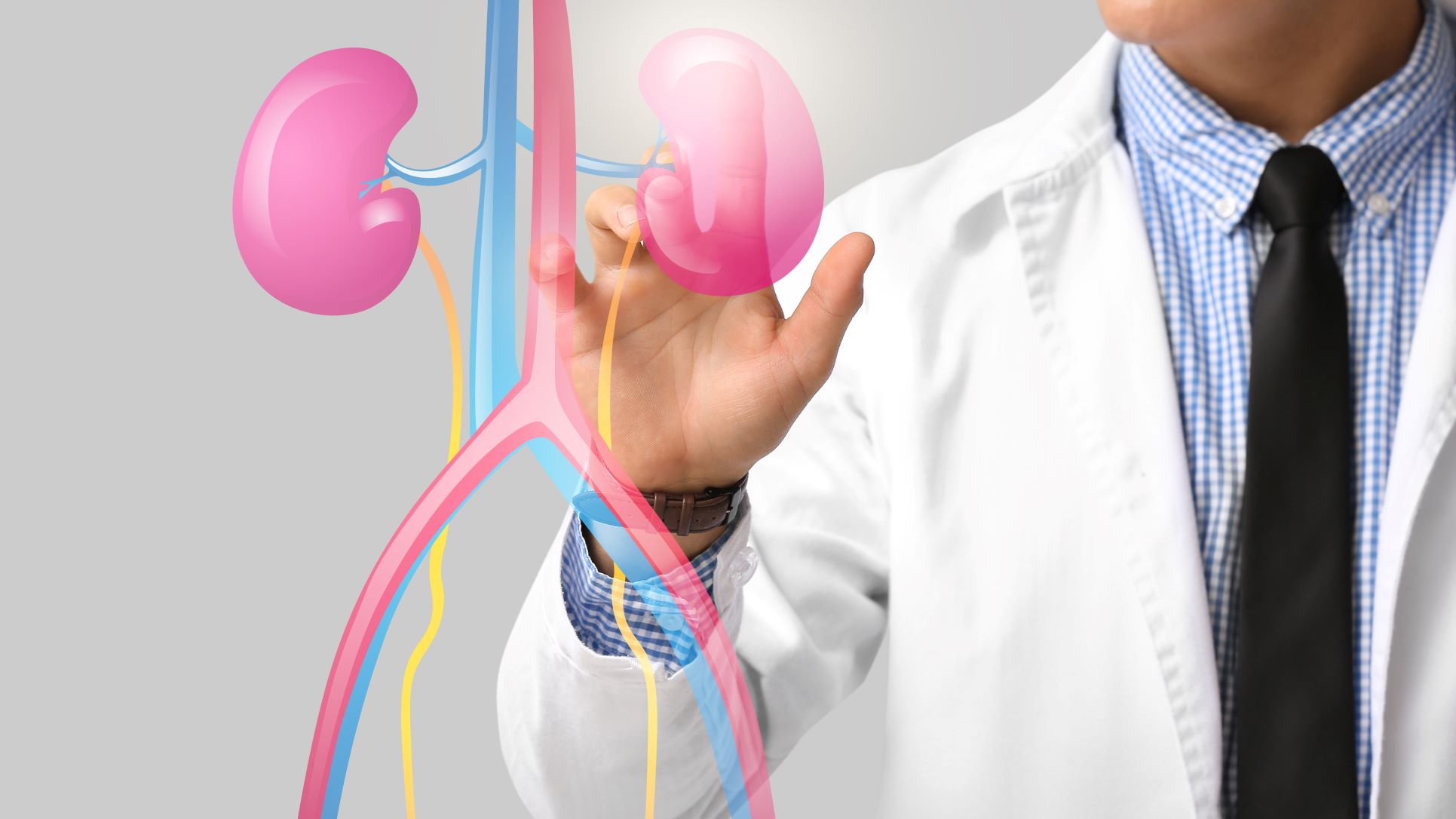
Renal lithotripsy refers to treatment methods used to break a kidney or urinary stone into smaller fragments so that the pieces can be removed or pass through the urinary tract.
The word lithotripsy essentially means stone fragmentation.
Depending on the location and characteristics of the stone, fragmentation can be achieved using externally generated shock waves or an energy source delivered directly to the stone through an endoscopic procedure.
The most commonly discussed approaches include:
-
Shock wave lithotripsy (SWL or ESWL)
-
Laser lithotripsy during ureteroscopy or flexible ureteroscopy
-
Other endoscopic stone-fragmentation techniques in selected procedures
The appropriate method depends on factors such as stone size, location, composition, density, anatomy, obstruction, infection and the patient's medical condition. Current European Association of Urology guidance lists SWL, retrograde intrarenal surgery and PCNL among the principal approaches for renal stones, with selection based on stone and patient characteristics.
At Prabhakar Bhurke Clinic, Dr. Sandip Prabhakar Bhurke, super specialist nephrologist and transplant physician, provides kidney-focused evaluation and can assess kidney function and related medical conditions when a patient has renal stones.
Why Is Lithotripsy Used for Kidney Stones?
Not every kidney stone requires immediate fragmentation.
Some small, uncomplicated stones can be monitored because spontaneous passage may be possible.
Active stone treatment may become appropriate when there is:
-
Persistent or recurrent pain
-
Urinary obstruction
-
Infection associated with the stone
-
Stone growth
-
Blood in the urine with relevant symptoms
-
Risk to kidney function
-
A stone unlikely to pass naturally
-
Other clinical or patient-specific reasons for removal
The decision is based on the complete clinical picture rather than the word “stone” on an imaging report.
The Main Types of Renal Lithotripsy
Shock Wave Lithotripsy
Shock wave lithotripsy (SWL), also called ESWL, uses externally generated shock waves to fragment the stone.
The patient does not need an instrument to be passed directly into the kidney for the fragmentation itself.
Once the stone has been broken into smaller pieces, the fragments can pass through the urinary tract.
NIDDK describes SWL as a procedure in which shock waves break a kidney stone into smaller pieces that can subsequently pass through the urinary tract.
Laser Lithotripsy
Laser lithotripsy is performed during an endoscopic procedure such as ureteroscopy or flexible ureteroscopy.
The specialist introduces a thin scope through the urinary tract and reaches the stone.
A laser is then used to fragment the stone into smaller pieces.
Depending on the situation, fragments may be extracted with a basket or reduced into small particles for passage.
EAU guidance supports holmium:YAG or thulium fiber laser lithotripsy for flexible ureteroscopy.
Lithotripsy During PCNL
For large or complex renal stones, fragmentation can also be performed through a percutaneous access route as part of percutaneous nephrolithotomy (PCNL).
A small tract is created through the skin into the kidney, allowing instruments to reach and remove or fragment the stone.
PCNL remains a standard approach for large renal calculi and is recommended as first-line treatment for stones larger than 2 cm when active treatment is indicated.
How Does Shock Wave Lithotripsy Work?
During SWL:
-
Imaging is used to locate the stone.
-
The patient is positioned so that the treatment system can focus shock waves on the stone.
-
Controlled shock waves are delivered.
-
The stone breaks into smaller fragments.
-
The fragments are allowed to pass naturally when appropriate.
-
Follow-up imaging may be performed to determine whether significant residual stone remains.
The effectiveness of SWL depends on stone size, location, composition, patient anatomy and other factors.
How Does Laser Lithotripsy Work?
Laser lithotripsy uses an endoscope to reach the stone directly.
A typical procedure involves:
-
Passing the ureteroscope through the natural urinary passage
-
Identifying the stone under direct vision
-
Applying laser energy to fragment the stone
-
Removing selected fragments with a basket when appropriate
-
Checking the urinary tract before completing the procedure
-
Placing a temporary ureteral stent when clinically indicated
NIDDK notes that ureteroscopy allows the doctor to visualize the urinary tract and remove or break stones, with many patients able to return home the same day depending on the procedure and clinical circumstances.
Which Renal Lithotripsy Method Is Suitable?
There is no single lithotripsy method that is appropriate for every kidney stone.
The treatment team may consider:
Stone Size
As stone size increases, SWL may become less effective and additional procedures may be needed.
For renal stones larger than 2 cm, EAU guidance recommends PCNL as the primary treatment option when active removal is indicated.
Stone Location
A stone in the renal pelvis may have different treatment considerations from a lower-pole calyceal stone.
Lower-pole anatomy can make fragment clearance more difficult after SWL in some patients.
Stone Composition and Density
Some stones are harder to fragment with shock waves.
CT stone density, often expressed in Hounsfield units, can provide information that helps with procedure selection.
Body Habitus and Stone-to-Skin Distance
Patient anatomy can influence whether shock waves can be effectively focused on the stone.
Urinary Obstruction
A stone causing obstruction may require active treatment rather than prolonged observation.
Infection
A urinary infection must be appropriately addressed before definitive stone removal.
An infected and obstructed urinary system can require urgent drainage rather than immediate definitive stone fragmentation.
Renal Lithotripsy Preparation
Before treatment, the specialist may review:
-
Ultrasound or CT findings
-
Stone size and location
-
Previous stone treatments
-
Kidney function
-
Urine examination
-
Urine culture when indicated
-
Current medications
-
Blood-thinning medicines
-
Other medical conditions
Imaging is particularly important because the stone's position and anatomy influence the treatment approach.
If a urinary infection is identified, it should be appropriately treated before planned stone removal.
Is Anaesthesia Required for Renal Lithotripsy?
The type of anaesthesia or sedation depends on the procedure and the patient's circumstances.
SWL may be performed with pain control, sedation or anaesthesia depending on the equipment, treatment setting and patient needs.
Ureteroscopy and laser lithotripsy generally require anaesthesia.
PCNL is also performed under anaesthesia.
The treating team will determine the safest approach after considering the patient's health and the planned procedure.
What Happens After Lithotripsy?
After stone fragmentation, the body may need to eliminate the remaining fragments.
Depending on the treatment, patients may experience:
-
Mild blood in the urine
-
Temporary urinary discomfort
-
Flank discomfort
-
Passage of stone fragments
-
Urinary frequency
-
Temporary stent-related symptoms
Follow-up imaging may be recommended to determine whether the stone has been completely cleared.
Residual fragments do not necessarily mean that the initial procedure failed, but significant remaining stone may require further management. EAU guidance notes that residual fragments can remain after SWL, ureteroscopy or PCNL and may require additional intervention in selected cases.
How Long Does Renal Lithotripsy Recovery Take?
Recovery depends on the treatment used.
Patients undergoing uncomplicated SWL may often return to normal activities relatively quickly, while recovery after PCNL can take longer.
After ureteroscopy with laser treatment, some patients experience urinary symptoms or discomfort for a short period, particularly if a stent has been placed.
Your treating specialist will provide individualized instructions regarding:
-
Activity
-
Fluid intake
-
Pain medicines
-
Antibiotics when indicated
-
Stent care
-
Follow-up imaging
-
Return to work or exercise
Do not assume that increasing water intake is appropriate for every patient, particularly when kidney function is impaired or fluid restriction has been prescribed.
What Are the Risks of Renal Lithotripsy?
As with any medical procedure, lithotripsy can have complications.
SWL-Related Risks
Possible complications include:
-
Blood in the urine
-
Pain
-
Residual stone fragments
-
Recurrent renal colic
-
Infection
-
Steinstrasse, in which multiple stone fragments accumulate in the ureter
-
Rare kidney or surrounding-tissue injury
EAU guidance reports that complication patterns vary according to patient and stone factors.
Ureteroscopy and Laser Risks
Potential complications include:
-
Urinary infection
-
Bleeding
-
Ureteral irritation or injury
-
Stent discomfort
-
Residual stone fragments
-
Need for another procedure
PCNL-Related Risks
PCNL is effective for large stones but is more invasive than SWL and may involve risks such as bleeding, infection and injury to surrounding structures.
The individual risk depends on stone complexity, anatomy, medical conditions and the procedure performed.
Can Renal Lithotripsy Remove a Large Kidney Stone?
It depends on the stone's size and location.
For some moderate-sized stones, SWL or flexible ureteroscopy may be suitable.
However, very large renal stones may be better treated with PCNL.
According to EAU guidance, renal stones larger than 2 cm should generally be treated primarily with PCNL when active removal is required because SWL may require multiple treatments and has a greater risk of obstruction from fragments.
Selected patients who cannot undergo PCNL may be considered for flexible ureteroscopy or SWL, although additional procedures may be necessary.
Can Lithotripsy Be Used for Lower-Pole Stones?
Yes, but the location requires special consideration.
Stone fragments produced by SWL must travel through the collecting system before leaving the kidney.
The anatomy of the lower pole can make fragment clearance less favorable in some cases.
For selected lower-pole stones, flexible ureteroscopy or PCNL may therefore be considered depending on stone size and other factors. EAU recommendations note that PCNL or retrograde intrarenal surgery can be considered for lower-pole stones when SWL effectiveness is limited.
What Is Steinstrasse After Lithotripsy?
Steinstrasse means an accumulation of stone fragments within the ureter following stone fragmentation, particularly after SWL.
The fragments can sometimes interfere with urine drainage and cause colic or obstruction.
Some cases can be managed conservatively, while others require additional treatment.
If fever or infection develops with obstruction, urgent decompression may be required.
When Is Lithotripsy Not Appropriate?
Lithotripsy may not be the right immediate treatment when:
-
There is an infected obstructed kidney
-
The stone is too large for effective SWL
-
Stone composition is unfavorable for shock-wave fragmentation
-
Anatomy makes stone targeting difficult
-
There is an untreated bleeding disorder
-
Another procedure offers a better chance of clearance
-
The patient's overall medical condition requires a different approach
The presence of infection with obstruction is particularly important. An obstructed kidney with signs of urinary infection or anuria is considered a urological emergency and generally requires urgent drainage.
Can Kidney Stones Be Dissolved Instead of Using Lithotripsy?
Most kidney stones cannot simply be dissolved with medication.
Certain uric acid stones may respond to medically supervised urine alkalinization.
For other stone types, physical removal or fragmentation may be necessary when active treatment is indicated.
Stone analysis and metabolic evaluation can help identify the stone type and guide future prevention.
How Can New Kidney Stones Be Prevented?
Removing a stone does not necessarily eliminate the reason it formed.
Depending on the stone type and individual risk factors, prevention may include:
-
Appropriate fluid intake
-
Reducing excessive dietary sodium
-
Maintaining a balanced diet
-
Avoiding unnecessary supplements
-
Stone analysis when available
-
Metabolic evaluation in recurrent stone formers
-
Appropriate preventive medication when prescribed
-
Regular follow-up for recurrent stone disease
The prevention plan should be individualized rather than based on a universal “kidney stone diet.”
When Should You Seek Urgent Care After Lithotripsy?
Contact your healthcare team promptly or seek urgent medical attention if you develop:
-
Fever or chills
-
Severe or worsening flank pain
-
Very little or no urine
-
Persistent vomiting
-
Severe weakness
-
Confusion
-
Heavy or persistent bleeding in urine
-
Severe difficulty passing urine
-
Rapid deterioration in your condition
Fever associated with urinary obstruction can indicate an infected obstructed system and requires urgent assessment.
Renal Stone Evaluation at Prabhakar Bhurke Clinic
At Prabhakar Bhurke Clinic, Dr. Sandip Prabhakar Bhurke, super specialist nephrologist and transplant physician, provides individualized kidney evaluation for patients with renal stones and associated kidney-function concerns.
Depending on the clinical situation, evaluation may include:
-
Kidney-function assessment
-
Creatinine and eGFR review
-
Urine examination
-
Infection assessment
-
Review of ultrasound or CT scans
-
Evaluation of urinary obstruction
-
Assessment of recurrent stone disease
-
Review of medical conditions and medications
-
Kidney-protection planning
-
Coordination with an appropriate urological or stone-treatment specialist when a procedure is required
The appropriate stone procedure depends on the imaging findings, stone characteristics and overall health of the patient.
Frequently Asked Questions
What is renal lithotripsy?
Renal lithotripsy is a group of procedures used to break kidney or urinary stones into smaller pieces so they can be removed or pass through the urinary tract.
Is renal lithotripsy the same as ESWL?
Not exactly. ESWL or SWL is one type of lithotripsy that uses externally generated shock waves. Lithotripsy can also be performed internally using laser energy during ureteroscopy or as part of other stone-removal procedures.
Is renal lithotripsy painful?
Discomfort varies according to the procedure. Pain-control medicines, sedation or anaesthesia may be used depending on the treatment and patient.
How many sessions of lithotripsy are required?
Some stones can be treated in one session, while others may require repeat treatment or a different procedure. Stone size, location, composition and the response to treatment influence the number of sessions.
Is laser lithotripsy better than shock wave lithotripsy?
The procedures have different advantages and limitations. The appropriate choice depends on stone size, location, density, anatomy, patient health and the likelihood of achieving adequate stone clearance.
Is lithotripsy suitable for a 2 cm kidney stone?
Treatment of a 2 cm stone requires individualized assessment. SWL, flexible ureteroscopy and PCNL may be considered depending on location and other factors. Stones larger than 2 cm are generally treated primarily with PCNL when active removal is indicated.
Can a kidney stone come back after lithotripsy?
Yes. Lithotripsy removes or fragments the existing stone but does not necessarily eliminate the metabolic or dietary factors responsible for stone formation. Preventive evaluation can reduce future risk.
Can I drink lots of water after lithotripsy?
Fluid intake should follow the instructions of your treating doctor. Excessive water is not appropriate for everyone, particularly people with kidney failure, heart disease or prescribed fluid restrictions.
Conclusion
Renal lithotripsy is an important part of modern kidney stone treatment, but it does not refer to just one procedure. Depending on the stone, treatment may involve shock wave lithotripsy, laser lithotripsy during ureteroscopy or stone fragmentation during PCNL.
The choice is influenced by the stone's size, location, density, composition, kidney anatomy, urinary obstruction, infection status and the patient's overall health. Large renal stones may require PCNL, while selected smaller stones can be treated with SWL or endoscopic techniques.
Successful stone care also involves follow-up after fragmentation and prevention of future stones. If a kidney stone is associated with fever, infection, obstruction, very low urine output or severe worsening symptoms, urgent medical assessment is required.
For kidney-function evaluation and individualized care related to renal stones, consult Dr. Sandip Prabhakar Bhurke at Prabhakar Bhurke Clinic.
Medical Disclaimer
This article is intended for general educational and informational purposes only and should not be considered a substitute for medical diagnosis, treatment or individualized medical advice. Renal lithotripsy includes different procedures, and the appropriate treatment depends on stone size, location, composition, density, urinary anatomy, obstruction, infection status and the patient's overall health. Not every patient is suitable for shock wave lithotripsy, laser lithotripsy or PCNL. Do not attempt to treat a kidney stone with excessive fluid intake, unprescribed medicines or herbal remedies. Fever, chills, severe flank pain, very little or no urine, persistent vomiting, confusion or sudden worsening after stone treatment may indicate a serious complication and requires urgent medical attention. Consult Dr. Sandip Prabhakar Bhurke or another qualified kidney or urinary-stone specialist for an individualized treatment plan.